Introduction
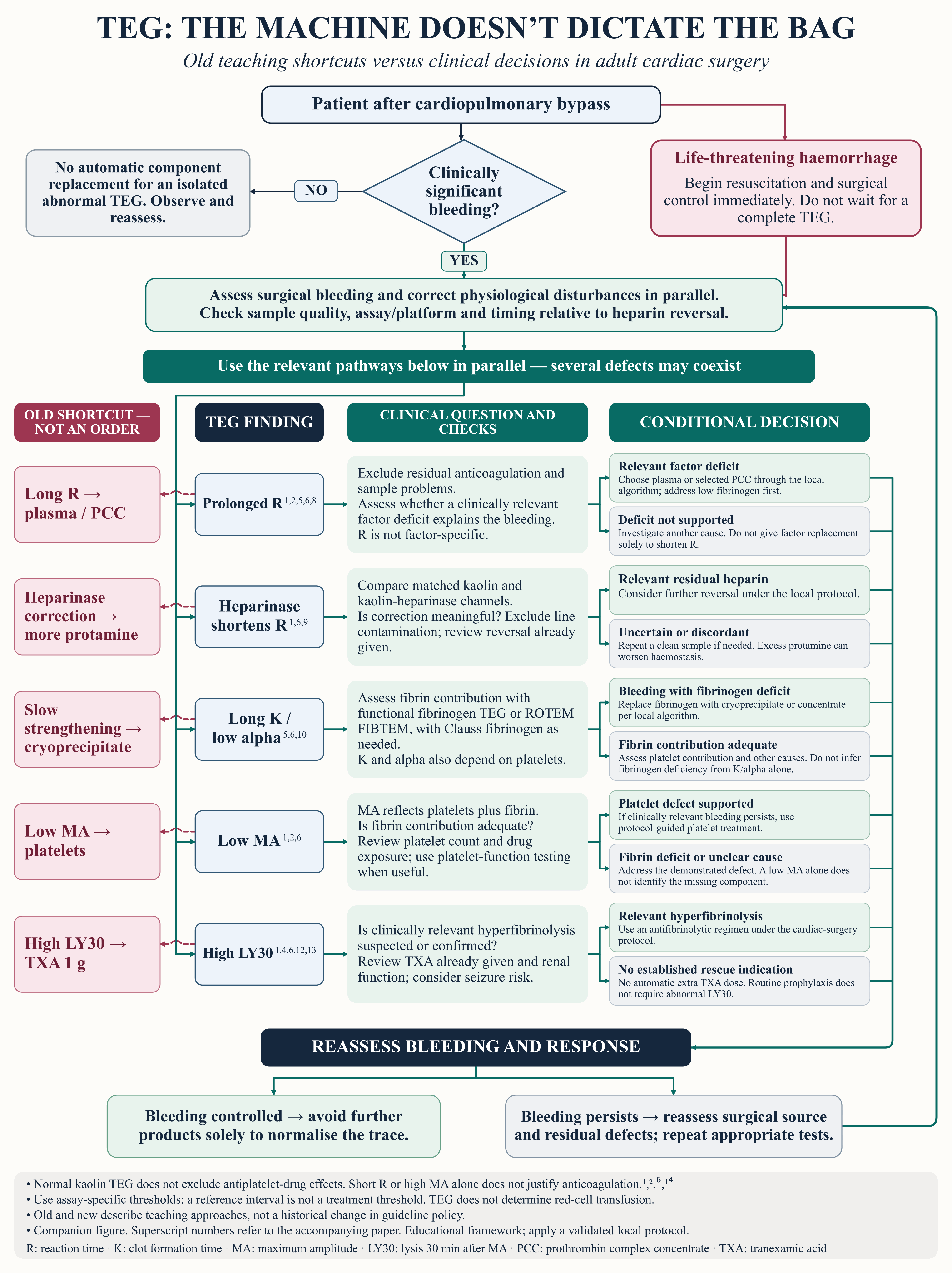
The familiar shorthand is easy to remember. A long reaction time means plasma; a low maximum amplitude means platelets. The difficulty begins when the association becomes an instruction. After cardiopulmonary bypass (CPB), a prolonged clotting time may reflect residual heparin rather than a factor deficiency, while a weak clot may arise from inadequate fibrin contribution even when the platelet count is acceptable. Hensley’s and Kaplan’s describe these overlapping mechanisms and the need to distinguish coagulopathic bleeding from a surgical source.1,2 A haemostatic product cannot close an unsecured vessel.
“Old” and “new” teaching are useful labels for the contrast between a product mnemonic and a clinical decision, but they should not imply that contextual interpretation was recently discovered. Hensley’s explicitly cautions against treating abnormal coagulation results without clinical justification. The 1999 trial by Shore-Lesserson and colleagues also evaluated a TEG-guided treatment algorithm, rather than the instrument in isolation.1,3 The problem is the loss of these qualifications when teaching is compressed into arrows between a tracing and a blood bag.
The 2021 STS/SCA/AmSECT/SABM guideline recommends goal-directed transfusion algorithms incorporating point-of-care testing, including viscoelastic devices, with a Class I, Level B-R recommendation. The 2024 European patient blood management guideline likewise supports algorithm-based haemostatic management.4,5 Neither endorsement makes a single abnormal TEG value a complete prescription. This review examines the common shortcuts and the evidence that should replace the missing diagnostic steps.
Scope of the review
This is a narrative review of adult cardiac surgery involving CPB. Relevant chapters from Hensley’s Practical Approach to Cardiothoracic Anesthesia and Kaplan’s Cardiac Anesthesia were examined alongside professional guidelines and selected clinical studies. Searches focused on the interpretation of TEG and on treatments commonly linked to its variables; the literature was checked through 30 July 2026. No systematic search protocol or formal risk-of-bias assessment was undertaken. Findings from adult cardiac surgery are not assumed to apply to paediatric practice or extracorporeal life support.
What the tracing can establish
Classical kaolin TEG describes clot development under specified assay conditions. Reaction time (R) ends when the tracing reaches an amplitude of 2 mm. K measures the subsequent time to 20 mm, whereas the alpha angle describes the rate of clot strengthening. Maximum amplitude (MA) reflects the strength of the platelet–fibrin clot. LY30 reports the percentage of clot lysis 30 minutes after MA, as calculated by the device. K cannot be determined if the tracing never reaches 20 mm. These measurements arise from the same evolving clot, so they cannot be read as independent assays of individual haemostatic components.6
The assay matters before any threshold is applied. Kaolin TEG and RapidTEG differ in activation, and functional fibrinogen assays answer a different question from the standard kaolin channel. ROTEM measurements are not numerical substitutes for TEG variables. Wong and colleagues found clinically relevant limits to interchangeability between TEG 5000 and TEG 6s in cardiac surgery, supporting the need for platform-specific algorithms.7
A reference interval is descriptive. It identifies the distribution of results in a reference population; it does not establish the point at which transfusion improves outcomes. For targeted treatment of post-CPB bleeding, the relevant question is whether an assay abnormality plausibly contributes to the bleeding and meets a locally validated intervention threshold. In a patient with satisfactory haemostasis, an isolated abnormal result usually does not justify component replacement. During life-threatening haemorrhage, however, treatment of a probable defect may have to begin before laboratory confirmation is available.1